
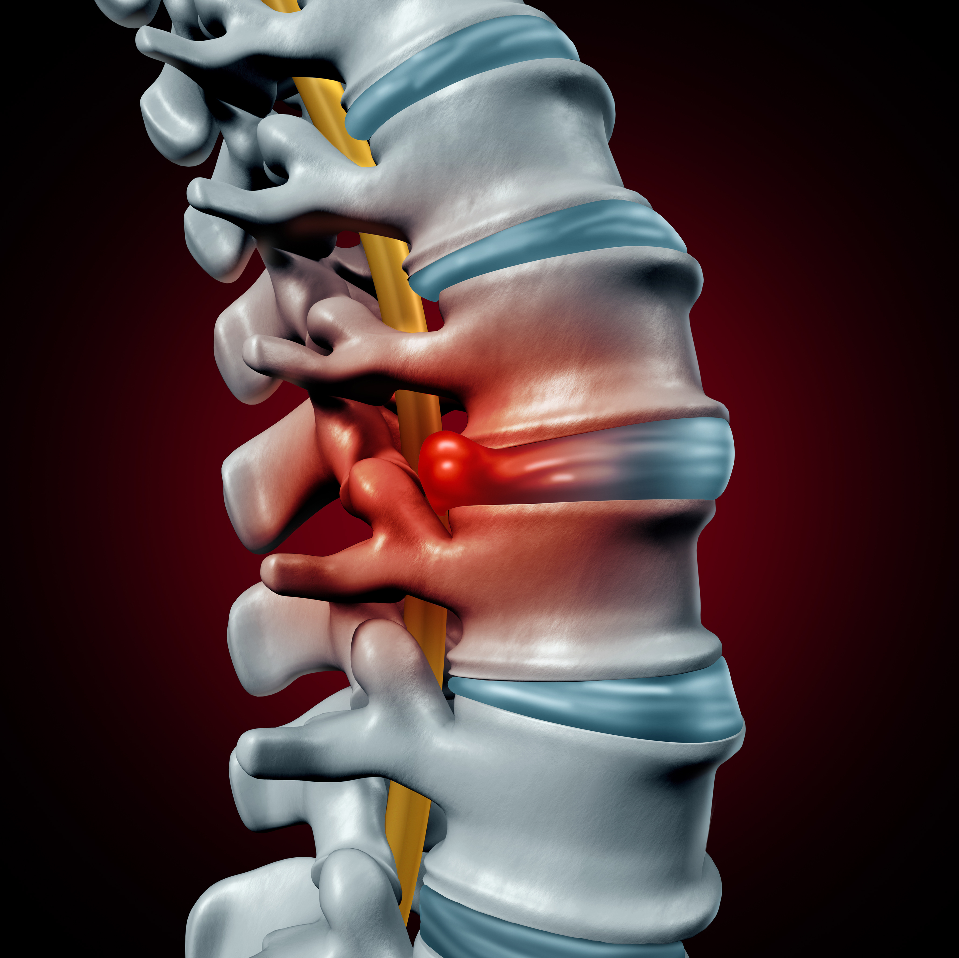
Lumbar canal stenosis or lumbar spine stenosis is a condition caused by reduction in the size of the canal, carrying nerves at the level of lumbar spine. This causes pressure on the nerves and hinder their functions.
Lumbar canal stenosis is quite frequent in the elderly and manifests as difficulty in walking.
What are the symptoms of Lumbar canal stenosis?
Lumbar stenosis is frequently asymptomatic and the correlation between the severity of symptoms and degree of stenosis of the spinal canal on radiology is poor.
Neurologic claudication is the most important symptom for this condition. Neurologic claudication means buttock or leg pain, or numbness that is worsened by walking or standing and relieved by rest(even standing). Sometimes, claudication in one leg can be confused with Sciatica.
Symptoms in the legs are usually bilateral. Patients can walk better with a stooped posture as this increases the spinal canal diameter. Usually patients do not have any significant muscle weakness or sensory deficits at rest in pure canal stenosis.
What are the causes of lumbar canal stenosis?
A few individuals may have congenitally or developmentally small canal. However, majority of patients have acquired lumbar canal stenosis. Certain factors that cause increased likelihood of tightness of canal include age related degeneration, trauma, and previous surgery. Certain medical conditions like epidural lipomatosis after steroid overuse, hypoparathyroidism, and renal dysfunction may also cause narrowing of the spinal canal.
How is Lumbar canal stenosis diagnosed?
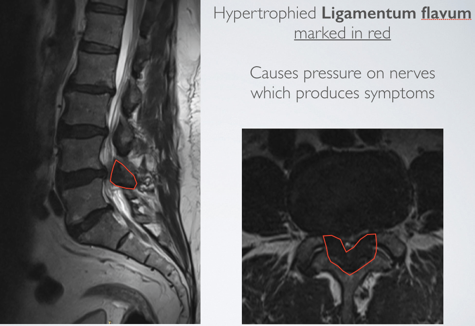
Lumbar canal stenosis is diagnosed based on history and clinical features primarily. It can be confirmed using an MRI of the Lumbar spine which will show reduction of the canal diameter and foramen for nerves. Dynamic x-rays of spine are also required to rule out instability, before making a final decision regarding treatment.
What are the treatment options of Lumbar canal stenosis?
Initially patients are offered non-surgical options which include exercise programs, spine stretching manoeuvres, epidural steroid injections and analgesics. Pain may improve over short periods, but long lasting pain relief in unlikely.
In patients with failed medical management or severe symptoms, surgical widening of the canal needs to be done.
This includes removal of bone forming the back side of spinal canal, and removing the ligamentum flavum which also becomes thick over time. The Lumbar spinal canal is decompressed and pressure is also relieved from the nerve roots. This procedure can now be routinely performed using keyhole approach with minimal blood loss and significantly shorter recovery times as compared to traditional open approach.
Surgery provides good relief in symptoms over long time, and patients feel the improvement in walking immediately after the surgery.
Patients may develop same level or adjacent level lumbar canal stenosis in 5-7 years after the surgery.
How is this lumbar canal stenosis surgery performed?
Conventionally the surgery for Lumbar canal stenosis was performed using a large muscle dissection to expose the lamina of the vertebra(open Lumbar laminectomy). The lamina was removed completely and the foramen through which the nerves exit the spinal canal would be widened. This would lead to a lot of scarring in the para-spinal muscles and significantly increase the post-op recovery.
With the current advances in spine surgery techniques, this decompression of the nerves can be achieved with much less trauma to the muscles. Using Key hole approach(also called MIS Lumbar decompression), through a small 1.5 cm incision, targeted bony removal and removal of ligament flavum can be achieved adequately to decompress the nerves. Upto three lumbar spinal segments can be decompressed using a single small incision, thereby significantly reducing the post-op pain and recovery time. Blood loss is minimal and most patients resume daily activities on first post-operative day.
For a single level, the surgery time is approximately 90 minutes, and the procedure is performed under General Anaesthesia.
The patient is positioned prone on the table in Operation theatre, and a C-arm is used to confirm the spinal level. After a small 1.5 cm incision, tubular dilators are passed and underlying bone is exposed(See video), and 14mm or 18mm working channel is introduced. Through this working channel bone is drilled using high speed drill and ligament flavum is excised. Part of facet joint between adjacent spinal segments is also removed to decompress the nerves. Incision is closed using absorbable sutures.
MIS Lumbar decompression
How is the recovery after MIS Lumbar decompression for Lumbar canal stenosis?
Minimally invasive spine decompression for lumbar canal stenosis is associated with very good results. Patients tend to feel the relief in leg pain and improvement in walking from first post-operative day itself. Patients can resume walking a few hours after the procedure and can be discharged next day after the surgery. Most patients go back to work and resume full activities within 2-3 weeks after the procedure.
Disclaimer- This is for the general awareness of the patients and cannot replace expert medical advice. Patient treatments need to be individualised and that can be decided based on clinical examination and evaluation by a trained physician.
Need an answer? Ask your question in the comment section below.

3 thoughts on “MIS Lumbar Decompression”